The recently published book “Tapeworms from the vertebrate bowels of the earth” edited by Drs. Janine Caira (EEB—UCONN) and Kirsten Jensen (EEB—University of Kansas) (see our previous post) was featured in UCONN Today with a gallery of stunning pictures of tapeworms, a group of organisms well represented in UCONN’s Biodiversity Research Collection.
Author: Bernard Goffinet
Army Ant video reaches milestone
 As part of the NSF funded project for the preservation of the Army Ant Guest Collection (AAGC), both videos created by Carl Rettenmeyer were shared with the public via Youtube.
This week, the documentary “Astonishing Ants” reached a milestone: the video has been viewed 10,000 times! The companion film “Associates of Eciton burchellii” has been viewed over 4200 times!
The video is widely praised by the viewers: “I learned 100x more in this video compared to a NatGeo doc” — “…refreshingly spartan” — “Fine documentaries like this should go viral” — “Possibly the best most in-depth doc i have ever seen on ants, particularly on army ants.thank u Dr.R. your hard work is appreciated”
For more news on the AAGC project, follow the project on Facebook.
New publication on mosses
 Medina R., M. Johnson, Y. Liu, N. Wilding, T. Hedderson, N. Wickett & B. Goffinet. 2018. Evolutionary dynamism in bryophytes: Phylogenomic inferences confirm rapid radiation in the family Funariaceae. Molecular Phylogenetics and Evolution 120: 240–247. pdf
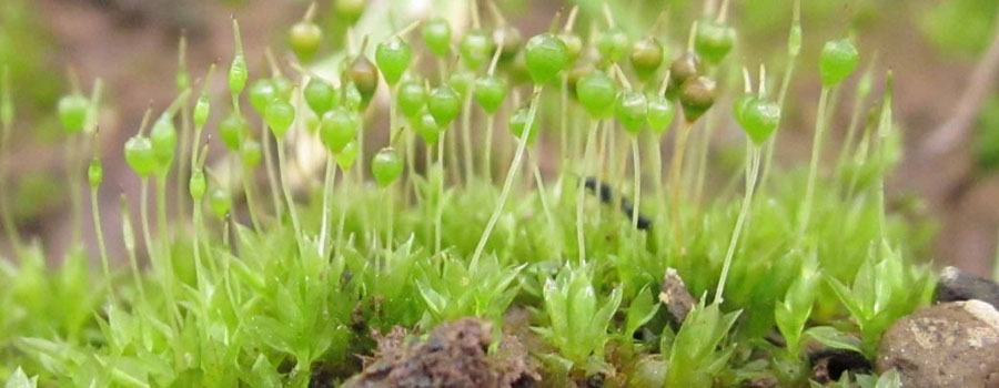
Abstract reads: Rapid diversifications of plants are primarily documented and studied in angiosperms, which are perceived as evolutionarily dynamic. Recent studies have, however, revealed that bryophytes have also undergone periods of rapid radiation. The speciose family Funariaceae, including the model taxon Physcomitrella patens, is one such lineage. Here, we infer relationships among major lineages within the Entosthodon-Physcomitrium complex from virtually complete organellar exomes (i.e., 123 genes) obtained through high throughput sequencing of genomic libraries enriched in these loci via targeted locus capture. Based on these extensive exonic data we (1) reconstructed a robust backbone topology of the Funariaceae, (2) confirmed the monophyly of Funariaand the polyphyly of Entosthodon, Physcomitrella, and Physcomitrium, and (3) argue for the occurrence of a rapid radiation within the Entosthodon-Physcomitrium complex that began 28 mya and gave rise more than half of the species diversity of the family. This diversification may have been triggered by a whole genome duplication and coincides with global Eocene cooling that continued through the Oligocene and Miocene. The Funariaceae join a growing list of bryophyte lineages whose history is marked by at least one burst of diversification, and our study thereby strengthens the view that bryophytes are evolutionarily dynamic lineages and that patterns and processes characterizing the evolution of angiosperms may be universal among land plants.
New publication on lichens
 Robert Lücking and colleagues completed a revision of a complex of lichen species, of which specimens from Chile, are deposited in CONN.
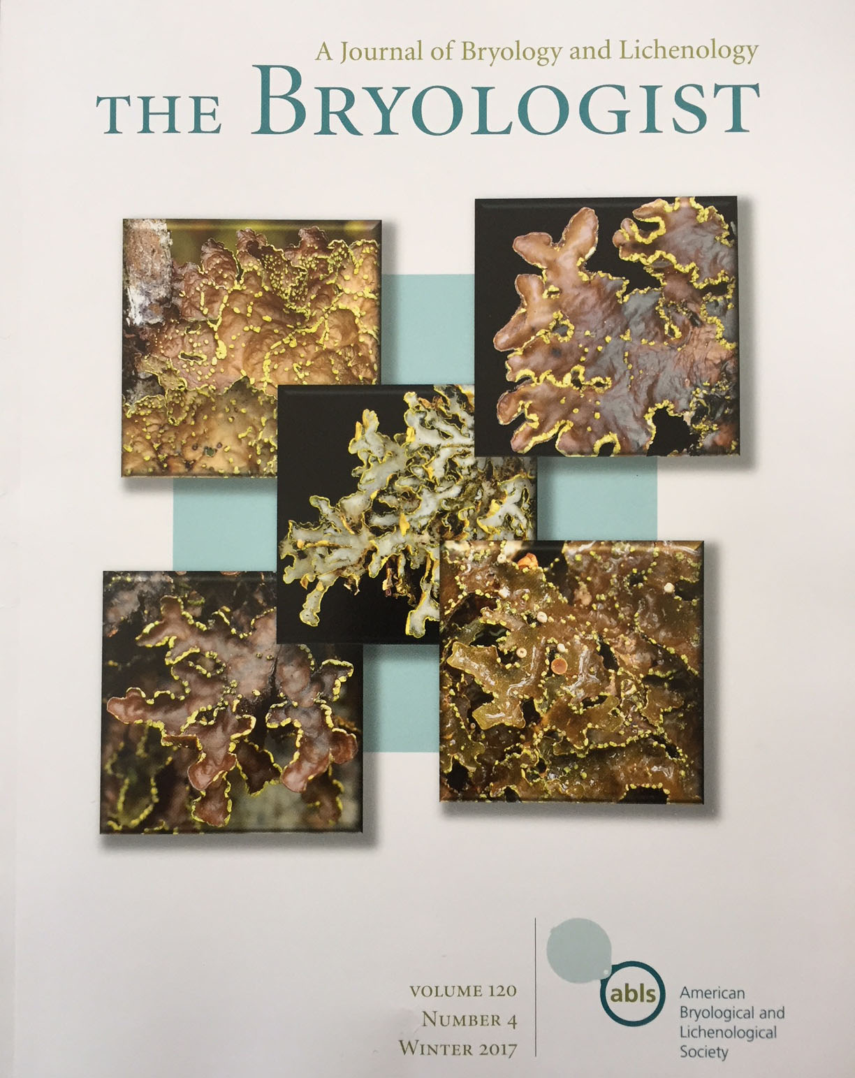
Lücking R., B. Moncada, B. McCune, E. Farkas, B. Goffinet, D. Parker, J. L. Chaves, L. Lőkös, P. R. Nelson, T. Spribille, S. Stenroos, T. Wheeler, A. Yanez-Ayabaca, K. Dillman, O. T. Gockman, T. Goward, J. Hollinger, E. A. Tripp, J. Villella, W. R. Álvaro-Alba, C. Julio Arango, M. E. S. Cáceres, L. Fernando Coca, C. Printzen, C. Rodríguez, K. Scharnagl, R. Rozzi, E. Soto-Medina & L. S. Yakovchenko. 2017. Pseudocyphellaria crocata (Ascomycota: Lobariaceae) in the Americas reveals to be ten species, and none of them is Pseudocyphellaria crocata. The Bryologist 120: 441–500.
Abstract reads: We provide a phylogenetic revision of the Pseudocyphellaria crocata complex in the Americas. Specimens traditionally identified as P. crocata, based on their cyanobacterial photobiont, yellow pseudocyphellae, at least partially white medulla, and yellow soralia or soralia-like structures, are shown to represent 13 distinct species, forming a monophyletic group divided into four large clades, three comprising one species each and one containing eight species, plus two taxa for which no molecular data are available. Seven species correspond to what was previously recognized as P. crocata and one to P. dozyana, whereas a further one is identified as the sorediate counterpart of the usually apotheciate taxon P. lechleri and another as a pseudosorediate morph of the usually phyllidiate species P. neglecta. Surprisingly, none of the species represents P. crocata s.str., which must therefore be excluded from the American lichen biota. The 13 recognized species include three species new to science and three new combinations: P. citrina (Gyeln.) Lücking, Moncada & S.Stenroos, comb. nov. [bas.: Cyanisticta citrina Gyeln., nom. nov. pro Sticta citrina Pers. nom. illeg.], P. desfontainii (Delise) Vain., P. deyi Lücking, sp. nov., P. dozyana (Mont. & Bosch) D.J.Galloway, P. epiflavoides (Gyeln.) Lücking, Farkas & Lokös, comb. nov. [bas.: Cyanisticta epiflavoides Gyeln.], P. hawaiiensis H.Magn., P. hillii (C.W.Dodge) D.J.Galloway, P. holarctica McCune, Lücking & Moncada, sp. nov., P. lechleri (Müll. Arg.) Du Rietz, P. neglecta (Müll. Arg.) H.Magn., P. punctata Lendemer, Lücking & Moncada sp. nov., P. sandwicensis (Zahlbr.) Moncada & Lücking, comb. nov. [bas.: Sticta crocata f. sandwicensis Zahlbr.], and P. xanthosticta (Pers.) Moncada & Lücking. Based on sequenced specimens, a neotype is selected for P. citrina and epitypes for P. hawaiiensis, P. lechleri, P. sandwicensis and P. xanthosticta. A key to all sorediate or pseudosorediate species of this complex in the Americas is presented, and all species are described, discussed and illustrated. pdf
New publication on lichens
A further contribution from the team working on the genus of lichenized fungi Peltigera, revealing 22 species new to science in a clade comprising 29 species total!
Magain N., J. Miadlikowska, O. Mueller, M. Gajdeczka, C. Truong, A. Salamov, I. Grigoriev, I. Dubchak, B. Goffinet, E. Sérusiaux & F. Lutzoni. Conserved genomic collinearity as a source of broadly applicable, fast evolving, markers to resolve species complexes: a case study using the lichen-forming genus Peltigera section Polydactylon. Molecular Phylogenetics and Evolution 117: 10–29. pdf
Abstract reads: Synteny can be maintained for certain genomic regions across broad phylogenetic groups. In these homologous genomic regions, sites that are under relaxed purifying selection, such as intergenic regions, could be used broadly as markers for population genetic and phylogenetic studies on species complexes. To explore the potential of this approach, we found 125 Collinear Orthologous Regions (COR) ranging from 1 to >10 kb across nine genomes representing the Lecanoromycetes and Eurotiomycetes (Pezizomycotina, Ascomycota). Twenty-six of these COR were found in all 24 eurotiomycete genomes surveyed for this study. Given the high abundance and availability of fungal genomes we believe this approach could be adopted for other large groups of fungi outside the Pezizomycotina. As a proof of concept, we selected three Collinear Orthologous Regions (COR1b, COR3, and COR16), based on synteny analyses of several genomes representing three classes of Ascomycota: Eurotiomycetes, Lecanoromycetes, and Lichinomycetes. COR16, for example, was found across these three classes of fungi. Here we compare the resolving power of these three new markers with five loci commonly used in phylogenetic studies of fungi, using section Polydactylon of the cyanolichen-forming genus Peltigera (Lecanoromycetes) – a clade with several challenging species complexes. Sequence data were subjected to three species discovery and two validating methods. COR markers substantially increased phylogenetic resolution and confidence, and highly contributed to species delimitation. The level of phylogenetic signal provided by each of the COR markers was higher than the commonly used fungal barcode ITS. High cryptic diversity was revealed by all methods. As redefined here, most species represent lineages that have relatively narrower, and more homogeneous biogeographical ranges than previously understood. The scabrosoid clade consists of ten species, seven of which are new. For the dolichorhizoid clade, twenty-two new species were discovered for a total of twenty-nine species in this clade.
New publication on mosses
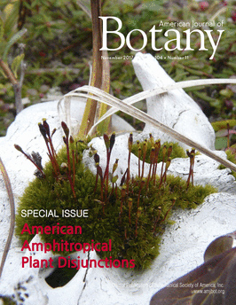
 Lily Lewis published the last paper from her dissertation as an invited paper in the Special November issue of the American Journal of Botany. All collections from Nepal, Alaska, Norway, Chile and Papua New Guinea are deposited in the CONN herbarium, and will serve further studies to assess species delimitations.
Lewis L.R., E.M. Biersma, S. Carey, K. Holzinger, S.F. McDaniel, R. Rozzi & B. Goffinet. Resolving the source region for the long-distance dispersal event that gave rise to the Magellanic sub-Antarctic endemic dung moss Tetraplodon fuegianus (Bryopsida: Splachnaceae). American Journal of Botany 104: 1651–1659. pdf
Abstract reads: PREMISE OF THE STUDY: American bipolar plant distributions characterize taxa at various taxonomic ranks but are most common in the bryophytes at infra-specifi c and infrageneric levels. A previous study on the bipolar disjunction in the dung moss genus Tetraplodon found that direct long-distance dispersal from North to South in the Miocene–Pleistocene accounted for the origin of the Southern American endemic Tetraplodon fuegianus , congruent with other molecular studies on bipolar bryophytes. The previous study, however, remained inconclusive regarding a specifi c northern hemisphere source region for the transequatorial dispersal event that gave rise to T. fuegianus .
METHODS: To estimate spatial genetic structure and phylogeographic relationships within the bipolar lineage of Tetraplodon , which includes T. fuegianus , we analyzed thousands of restriction-site-associated DNA (RADseq) loci and single nucleotide polymorphisms using Bayesian individual assignment and maximum likelihood and coalescent model based phylogenetic approaches.
KEY RESULTS: Northwestern North America is the most likely source of the recent ancestor to T. fuegianus.
CONCLUSIONS: Tetraplodon fuegianus , which marks the southernmost populations in the bipolar lineage of Tetraplodon , arose following a single long-distance dispersal event involving a T. mnioides lineage that is now rare in the northern hemisphere and potentially restricted to the Pacifi c Northwest of North America. Furthermore, gene fl ow between sympatric lineages of Tetraplodon mnioides in the northern hemisphere is limited, possibly due to high rates of selfi ng or reproductive isolation.
New publications on plants
Two new publications from the Les lab on Elatine, a genus of aquatic plants:
1. Razifard, H., D. H. Les & G. C. Tucker. 2017. Reticulate evolution in Elatine L.(Elatinaceae), a predominantly autogamous genus of aquatic plants. Systematic Botany 42: 87-95. pdf
Abstract reads: The study of hybridization in aquatic plants is complicated by rarity of flower production, absence of roots, and asexuality. Elatine is a cosmopolitan genus of aquatic flowering plants with about 25 species worldwide. Historically, there has been little concern regarding hybridization in the genus due to the prevalence of autogamy (i.e. self-pollination), which potentially limits xenogamous pollen transfer among the species. Two morphologically complex species (Elatine hexandra and E. americana) are the only known polyploids in the genus. In previous phylogenetic analyses, both species resolved incongruently in gene trees obtained from nuclear (ITS) versus plastid (matK/trnK and rbcL) regions. Suspecting that the phylogenetic incongruence might be a consequence of past hybridization events, we tested that hypothesis by conducting an additional phylogenetic analysis of Elatine, which incorporated sequences from a low copy nuclear gene (phyC). Elatine hexandra and E. americana were the only Elatine species exhibiting intraspecific polymorphic sites, i.e. heterozygosity, in phyC. Allele specific amplification enabled us to resolve these polymorphisms for inclusion in a phylogenetic analysis along with the monomorphic phyCsequences within species obtained for the remaining Elatine species. The phyC tree confirmed that both polyploids probably are allopolyploids, in a pattern consistent with the placement of the putative parental taxa in previous phylogenetic analyses of ITS, matK/trnK,and rbcL sequence data. The distributions of E. americana and E. hexandra, along with their potential parental species, are consistent with the proposed hybrid origins for the polyploids and provide additional clues on their geographic regions of origin.
2. Razifard, H., A. J. Rosman, G. C. Tucker & D. H. Les. 2017. Systematics of the cosmopolitan aquatic genus Elatine. Systematic Botany 42: 73–86. pdf
Abstract reads: The cosmopolitan genus Elatine (Elatinaceae) includes about 25 aquatic species of mostly diminutive aquatic plants, whose relationships have not been evaluated using a phylogenetic approach. The taxonomic study of this group has been complicated by the small stature of the plants, their minute reproductive structures, and their cosmopolitan distribution. Consequently, much uncertainty exists with respect to species delimitations, their geographical distributions, and interspecific relationships. To clarify the infrageneric classification of Elatine and to provide insights on interspecific relationships within the genus, we conducted a phylogenetic study of nearly all (24) of the currently recognized species using both morphological and molecular data. The tree topology obtained based on morphological data (including vegetative and reproductive characters) was less-resolved than the trees based on molecular data, derived from either nuclear (ITS) or two plastid regions (matK/trnK and rbcL). However, the tree topology obtained from combined morphological and molecular data was well resolved and placed the morphologically distinctive E. alsinastrum as the sister group of the remaining species, which fell within two major clades: a clade of 4-merous-flowered species and a clade of 3-merous species, within which was embedded a subclade of 2-merous species. Although a number of topological differences occurred between the ITS and plastid tree topologies, significant incongruence was observed only for the placements of E. americana and E. hexandra, possibly resulting from reticulate evolution. Bergia, the sister genus of Elatine, comprises larger species, which often are mostly helophytic but never truly aquatic. Ancestral state reconstructions based on the ITS tree indicated that a morphological reduction series (in stature and floral merosity) exists among Elatine species, which is best explained as a consequence of adaptation to their aquatic life. These phylogenetic analyses also have helped to clarify the infrageneric classification of the genus and to provide a better understanding of the natural and nonindigenous distributions of the species. The new monotypic section Elatine sect. Cymifera , including E. brochonii, is described.
New publications on parasites
Three new studies were published by the Caira lab in EEB that further highlight the diversity and evolution of parasites.
1. Bueno V. M. & J. N. Caira. 2017. Redescription and molecular assessment of relationships among three species of Echeneibothrium (Rhinebothriidea: Echeneibothriidae) parasitizing the Yellownose Skate, Dipturus chilensis, in Chile. Journal of Parasitology 103: 268–284. pdf
Abstract reads: Much progress has recently been made in revising the taxonomic assignments of genera originally classified in the polyphyletic “Tetraphyllidea.” Many of these genera, including Echeneibothrium, were accommodated in the order Rhinebothriidea. However, beyond this larger taxonomic action, little work has been conducted on this genus over the past 50 yr. Consequently, the criteria used for characterizing species of Echeneibothrium have lagged behind those typically used in more modern descriptions of elasmobranch-hosted cestode taxa. A series of collecting trips to Chile to obtain cestodes from the yellownose skate, Dipturus chilensis, provided a unique opportunity to apply modern morphological and molecular methods to investigate the 3 species of Echeneibothrium reported parasitizing this skate, specifically Echeneibothrium megalosoma, Echeneibothrium multiloculatum, and Echeneibothrium williamsi. In addition to redescribing all 3 species, using morphological data from light and scanning electron microscopy, maximum likelihood and bayesian inference phylogenetic analyses of the D1–D3 regions of the 28S rDNA gene were conducted to assess their relationships among other echeneibothriids for which comparable data are available. Sequencing of 59 specimens representing these 3 species of Echeneibothrium allowed us to assess the intra- and interspecific variation in the 28S rDNA gene. The redescriptions use standardized terminology for scolex morphology, proglottid anatomy, and microthrix forms and pattern; they also expand on the original descriptions to include data on scolex size, ovary size, vas deferens and vaginal configurations, testes arrangement, and genital pore position. Our morphological work led to a major reinterpretation of the scolex morphology with the recognition that all 3 species bear an apical bothridial sucker, rather than an apical loculus, prompting emendation of the diagnosis for the family Echeneibothriidae. The presence of a band of spinitriches at the apex of the apical modification of the scolex proper seems to represent an important feature for distinguishing the 2 portions of the myzorhynchus across species. Intraspecific variation ranged from 0 to 7 bp across species and interspecific variation ranged from a low of 39–46 bp between E. williamsi and E. multiloculatum to a high of 61–66 bp between E. multiloculatum and E. megalosoma. Phylogenetic analyses indicate that the 3 species of Echeneibothrium hosted by the yellownose skate are not each other’s closest relatives, suggesting multiple colonization events of D. chilensis have occurred. Further phylogenetic investigation is also likely to confirm the status of the genus Pseudanthobothrium as a synonym of Echeneibothrium because its species generally group among members of Echeneibothrium.
2. Caira, J. N., C. J. Healy, F. P. Marques & K. Jensen. 2017. Three new genera of rhinebothriidean cestodes from stingrays in Southeast Asia. Folia parasitologica 64: 008. pdf
Abstract reads: Three genera of rhinebothriideans, previously referred to as New genus 1, New genus 2 and New genus 4, are erected in the the Anthocephaliidae. New genus 1 is established as Divaricobothrium gen. n., with Divaricobothrium tribelum sp. n. as its type species; Echeneibothrium trifidum Shipley et Hornell, 1906 is transferred to the genus as Divaricobothrium trifidum (Shipley et Hornell, 1906) comb. n. This genus is unique among rhinebothriidean genera in bearing bothridia that are posteriorly deeply divided into two lobes with facial loculi but no apical sucker, and a vagina that extends to near the anterior margin of the proglottid. Its species parasitise Indo-Pacific members of the genera Brevitrygon Last, Naylor et Manjaji-Matsumoto, Maculabatis Last, Naylor et Manjaji-Matsumoto and Pateobatis Last, Naylor et Manjaji-Matsumoto. New genus 2 is established as Barbeaucestus gen. n., with Barbeaucestus jockuschae sp. n. as its type species; Barbeaucestus ralickiae sp. n. is also described. Anthobothrium sexorchidum Williams, 1964 and Rhinebothrium shipleyi Southwell, 1912 are transferred to the genus as Barbeaucestus sexorchidus (Williams, 1964) comb. n. and Barbeaucestus shipleyi (Southwell, 1912) comb. n., respectively. This genus is unique among rhinebothriidean genera in that its bothridia are substantially wider than long, bear an apical sucker and at least one row of two or more facial loculi in their anterior half. Its species parasitise the genera Neotrygon Castelnau and Taeniura Müller et Henle. New genus 4 is established as Sungaicestus gen. n. with transfer of Rhinebothrium kinabatanganensis Healy, 2006, as Sungaicestus kinabatanganensis (Healy, 2006) comb. n., as its type species. Among the genera of its order, this genus most closely resembles Rhinebothrium Linton, 1890, however, despite the original description, the bothridia were found to bear, rather than lack, apical suckers. This monotypic genus is known only from the freshwater stingray Urogymnus polylepis (Müller et Henle). The familial diagnosis of the Anthocephaliidae Ruhnke, Caira et Cox, 2015 is emended. The family now houses five genera.
3. Gallagher K., J. N. Caira & M. Cantino. 2017. Ultrastructure of cell types within the scolex and cephalic peduncle of the litobothriidean tapeworm, Litobothrium aenigmaticum. Invertebrate Biology 136: 243–259. pdf
Abstract reads: Litobothrium aenigmaticum Caira, Jensen, Waeschenbach, & Littlewood 2014 is a cestode species that parasitizes pelagic thresher sharks in Taiwan and the Gulf of California. A previous study using light microscopy suggested the scolex contains four types of unusual tissues, one of which was considered to aid in adhesion of the worm to the mucosa of the spiral intestine. The function of the other tissues was unknown. Also unknown was the function of two laterally paired ducts found extending throughout the length of the worm. The goal of the present study was to use transmission electron microscopy (TEM) to examine the internal anatomy of the scolex and cephalic peduncle of this tapeworm in more detail. Three specimens from Taiwan were examined with TEM and two with light microscopy. Transmission electron microscopy revealed that the laterally paired ducts were excretory, since they were closely associated with numerous protonephridia. The number of cell types, previously referred to as tissues, in the scolex was expanded to 11 based on a combination of features including cell size, nucleus size, ratio of the maximum diameter of the nucleus to maximum diameter of the whole cell, and types of observed organelles, as well as the presence or absence of cytoplasmic electron-dense and electron-lucent vesicles. The cytoplasm of all 11 cell types was found to include electron-dense vesicles, and all types were periodic acid-Schiff (PAS) positive. This suggests that the electron-dense vesicles may contain glycoproteins and/or mucoproteins. As all 11 cell types occurred within the region of the scolex that is surrounded by a papilla of host mucosa at the site of tapeworm attachment, we hypothesize that one or more of these cell types may produce a substance that induces this inflammatory reaction. The specific products in the secretory vesicles and how those products are released are still unclear, as are any relationships between or among the cell types in terms of products produced. Further research will be necessary to fully understand the complexity of this organism.
New publication on algae
Simona Augyte who completed her Ph.D. dissertation in EEB with Dr. C. Yarish, lead-authored a publication focused on the brown alga Saccharina angustissima. Specimens studied are deposited in the CONN herbarium. The reference of the publication is: Augyte, S., L. Lewis, S. Lin, C. D. Neefus and C. Yarish. 2018. Speciation in the exposed intertidal zone: the case of Saccharina angustissima comb. nov. & stat. nov. (Laminariales, Phaeophyceae). Phycologia 57: 100–112. pdf
The abstract reads: Saccharina latissima is a perennial kelp with a circumboreal distribution from the North Pacific to the North Atlantic coasts. Our study clarified the taxonomy of the morphologically distinct Saccharina latissima forma angustissima (Collins) A.Mathieson from the low intertidal zone on exposed islands and ledges of Casco Bay, Maine, USA. To identify genetic divergence between the two morphotypes, S. latissima and S. latissima f. angustissima, we used a multilocus phylogenetic approach including nuclear-encoded internal transcribed spacer, mitochondrial cox1 and cox3, and plastid-encoded rbcL gene sequences. Genetic analysis of the individual markers and combined data set using SVDquartets resulted in p-distance values for all markers of , 1%, suggesting low divergence between the two forms. However, there was as much or more genetic divergence between S. latissima and S. latissima f. angustissima as there were between other taxonomically accepted species of Saccharina. To investigate sexual compatibility between the two forms, we made reciprocal crosses of the gametophytes and observed sporophyte formation. All crosses were successfully grown to the juvenile sporophyte stage, suggesting that the two are reproductively compatible in vitro. It is unknown if the two populations freely hybridize in the field. Last, we compared wave action, the ecological factor most likely driving the unique morphology, at exposed sites with S. latissima f. angustissima and protected sites with S. latissima. The mean wave force at the exposed site was over 30 times higher in magnitude than at the protected site at 160.04 6 32.58 N and 4.75 6 6.75 N, respectively, during the summer. The significant differences in morphology, the lack of specimens with intermediate morphologies, and the results of a common garden experiment suggest that the morphological differences in S. latissima f. angustissima are heritable with a genetic basis. Therefore, on the basis of our molecular evidence coupled with ecological studies, we are elevating S. latissima f. angustissima (Collins) A.Mathieson to specific rank as S. angustissima (Collins) Augyte, Yarish & Neefus comb. nov.& stat. nov.
Biases in herbarium collections
Dr. Daru (Harvard) and collaborators are presenting a study of holdings of herbaria in three regions with complete digitized floras, including New England, and thus based on CONN specimens. Their study, entitled “Widespread sampling biases in herbaria revealed from large-scale digitization” is to appear in the New Phytologist.
The summary reads:
- Nonrandom collecting practices may bias conclusions drawn from analyses of herbarium records. Recent efforts to fully digitize and mobilize regional floras online offer a timely opportunity to assess commonalities and differences in herbarium sampling biases.
- We determined spatial, temporal, trait, phylogenetic, and collector biases in c. 5 million herbarium records, representing three of the most complete digitized floras of the world: Australia (AU), South Africa (SA), and New England, USA (NE).
- We identified numerous shared and unique biases among these regions. Shared biases included specimens collected close to roads and herbaria; specimens collected more frequently during biological spring and summer; specimens of threatened species collected less frequently; and specimens of close relatives collected in similar numbers. Regional differences included overrepresentation of graminoids in SA and AU and of annuals in AU; and peak collection during the 1910s in NE, 1980s in SA, and 1990s in AU. Finally, in all regions, a disproportionately large percentage of specimens were collected by very few individuals. We hypothesize that these mega-collectors, with their associated preferences and idiosyncrasies, shaped patterns of collection bias via ‘founder effects’.
- Studies using herbarium collections should account for sampling biases, and future collecting efforts should avoid compounding these biases to the extent possible.